
A new co-training AI algorithm for medical imaging that can effectively mimic the process of seeking a second opinion has been designed by researchers at Monash University.
Artificial Intelligence is revolutionising the way diagnostic imaging is performed, offering a new level of accuracy, efficiency, and patient experience.
In 2023, AI in radiology is poised to make a big impact in the medical field, offering innovative solutions that have the potential to transform the entire diagnostic imaging industry.
This research, by Monash University faculties of Engineering and IT, will advance the field of medical image analysis for radiologists and other health experts.
Published recently in Nature Machine Intelligence, the research addressed the limited availability of human annotated, or labelled, medical images by using an adversarial, or competitive, learning approach against unlabelled data.
Read More

PhD candidate Himashi Peiris of the Faculty of Engineering, said the research design had set out to create a competition between the two components of a “dual-view” AI system.
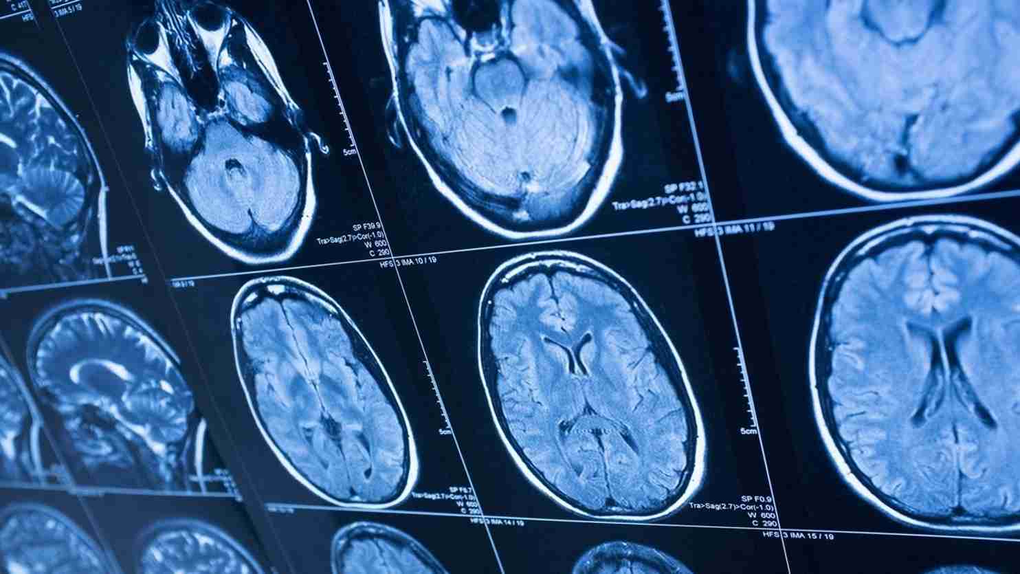
AI algorithms for medical imaging can leverage deep learning and machine learning techniques to analyse vast amounts of medical data, such as X-rays, CT scans, MRIs, and ultrasounds.
By harnessing this technology’s potential, medical practitioners gain an unprecedented ability to detect and identify anomalies, even in the earliest stages of diseases, considerably improving patient outcomes.
“One part of the AI system tries to mimic how radiologists read medical images by labelling them, while the other part of the system judges the quality of the AI-generated labelled scans by benchmarking them against the limited labelled scans provided by radiologists,” said Ms Peiris.
“Traditionally radiologists and other medical experts annotate, or label, medical scans by hand highlighting specific areas of interest, such as tumours or other lesions. These labels provide guidance or supervision for training AI models.
“This method relies on the subjective interpretation of individuals, is time-consuming and prone to errors and extended waiting periods for patients seeking treatments.”
The availability of large-scale annotated medical image datasets is often limited, as it requires significant effort, time and expertise to annotate many images manually.
The algorithm developed by the Monash researchers allows multiple AI models to leverage the unique advantages of labelled and unlabelled data, and learn from each other’s predictions to help improve overall accuracy.
“Across the three publicly accessible medical datasets, utilising a 10 per cent labelled data setting, we achieved an average improvement of 3 per cent compared to the most recent state-of-the-art approach under identical conditions,” said Ms Peiris.
“Our algorithm has produced groundbreaking results in semi-supervised learning, surpassing previous state-of-the-art methods. It demonstrates remarkable performance even with limited annotations, unlike algorithms that rely on large volumes of annotated data.
“This enables AI models to make more informed decisions, validate their initial assessments, and uncover more accurate diagnoses and treatment decisions.”
The Applications of AI In Medical Imaging
The applications of AI in medical imaging extend to various medical specialties, including oncology, cardiology, neurology, and orthopedics.
For instance, in oncology, AI algorithms can assist in early tumor detection and tracking treatment response, enabling oncologists to devise tailored therapeutic strategies for cancer patients.
In cardiology, AI can aid in detecting subtle cardiac abnormalities, contributing to the prevention of cardiovascular diseases. In neurology, AI algorithms can facilitate the identification of neurological disorders and assist in predicting disease progression.
Additionally, in orthopedics, AI can provide precise assessments of musculoskeletal conditions, leading to more accurate surgical planning and better patient outcomes.
The next phase of the research will focus on expanding the application to work with different types of medical images and developing a dedicated end-to-end product that radiologists can use in their practices.